Retinal Clinic

Advanced Retina Services at Anil Eye Hospital

At Anil Eye Hospital, our state-of-the-art Retina Department is dedicated to diagnosing and treating complex retinal conditions using the latest technology and a specialist-led approach. Whether you’re facing diabetic eye issues or sudden vision loss, our experienced retina specialists offer precise, compassionate care tailored to you.

Inside Our Retina Department

Our retina unit is one of the most advanced in Thane district, staffed with highly trained retina surgeons and supported by modern infrastructure.

Our Retina Department Includes:

  • Green Laser for diabetic retinopathy and retinal tears
  • OCT (Optical Coherence Tomography) for detailed retinal imaging
  • Fundus Fluorescein Angiography (FFA) for retina blood flow analysis
  • Vitrectomy System for advanced retinal surgeries
  • Intravitreal Injection facilities for age-related macular degeneration (AMD), diabetic macular edema, and retinal vein occlusions
  • Digital Fundus Photography for retina documentation
  • Full-time retina specialists and retina-trained optometrists

Types of LASIK & Vision Correction Procedures

Diabetic Retinopathy

Diabetic retinopathy is a health condition that occurs in diabetic people as a result of damage to the retina’s blood vessels. Diabetic retinopathy can develop if a person has type 1 or 2 diabetes and a long history of uncontrolled high blood sugar levels.

Stages of Diabetic Retinopathy

  1. Non-proliferative Diabetic Retinopathy (NDPR)– Also called ‘early diabetic retinopathy’
  2. Proliferative Diabetic Retinopathy (PDR)

Symptoms of Diabetic Retinopathy
• seeing floaters or dark spots
• objects appear bent or slanting (metamorphosis)
• blurred vision
• sudden loss of vision
• difficulty distinguishing colors

Diagnosis and treatment of DR:

A doctor can diagnose diabetic retinopathy by doing a dilated eye exam. They may also perform a fluorescein angiography test, Optical coherence tomography (OCT) exam. Early diagnosis and periodic follow-ups are key to this condition. 

At AEH, Thane, retina specialists screen all the diabetic patients, which includes DFE & various scans & provide the treatment as earlier as possible.

Macular Degeneration

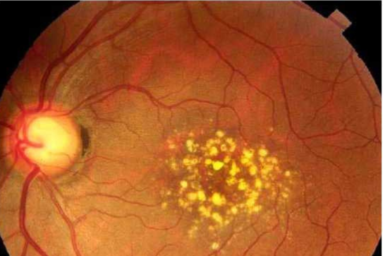
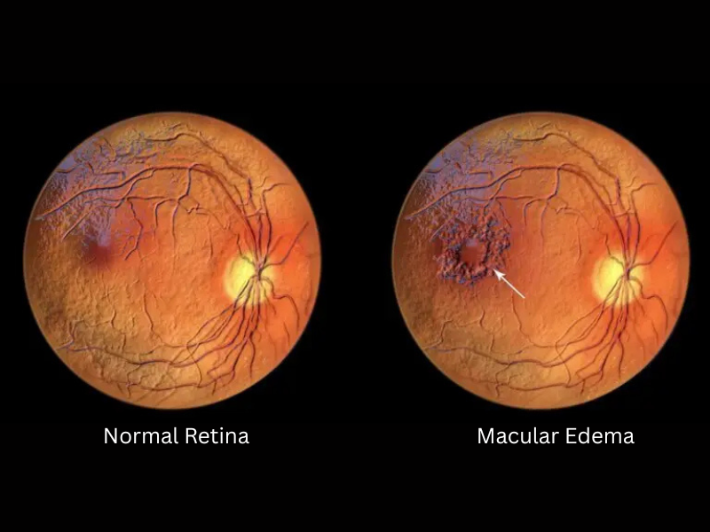
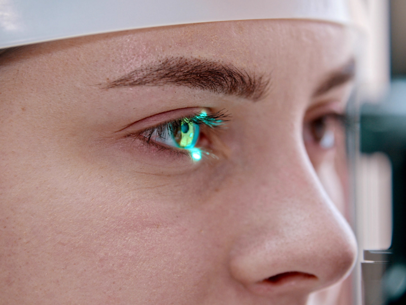
ARMD or AMD is caused by deterioration of the central part of the retina. The condition often does not have any symptoms, to begin with, but with progression, it may cause severe visual impairment, even complete blindness( in some cases).

Dry Macular Degeneration vs. Wet Macular Degeneration

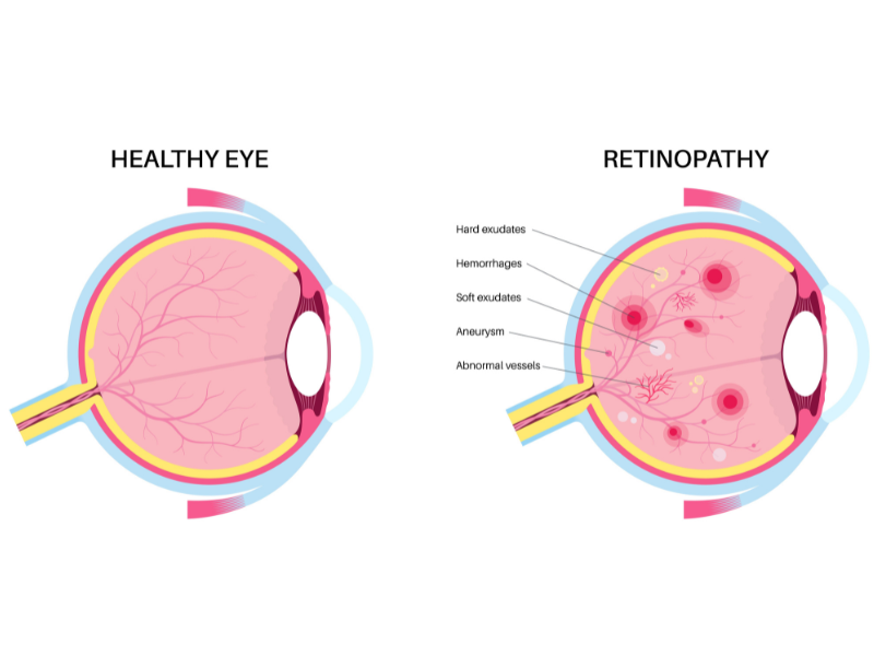
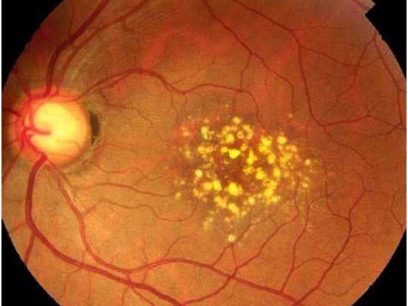
In Dry Macular Degeneration, yellow deposit is deposited at the macula which cause impairment in vision with growth in size or increase in number.

On the other hand, in Wet Macular Degeneration, leaky abnormal blood vessels and fluid into the retina. This results in loss of central vision.

Those highest at risk for age-related macular degeneration are over the age of 55, smokers, or those with a family history of AMD.

The loss of central vision in AMD can interfere with simple everyday activities, such as the ability to see faces, drive, read, write, or do close work, such as cooking or fixing things around the house.

There are ways to slow its effects. It is possible to lower the risk of AMD or slow its progression by eating healthy, exercising, and protecting your eyes from ultraviolet light.

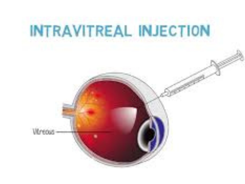
If you are above 50 years of age & experience any difficulties in central vision, you can visit AEH, Thane where retina specialist will not only help with the diagnosis & but also help you to lower the progression of AMD through various techniques like intravitreal antiVEGFs.

Retinopathy of Prematurity

When a baby is born more than two months prematurely, there is an increased chance they will be born with a condition known as retinopathy of prematurity (ROP), which is characterized by the growth of abnormal blood vessels in the fetal retina.

Over time, the effects of ROP may lead to:

  • Retina damage or detachment, causing permanent vision loss
  • Nearsightedness, or myopia
  • Amblyopia, also known as a “lazy eye”
  • Crossed eyes, or strabismus

When babies are born prematurely, they are sent to the hospital’s neonatal intensive care unit, or NICU (NICU), where they undergo testing to quantify their overall health. This usually includes an eye exam to determine if there are any vision issues or retinal problems. The ophthalmologist will put eye drops in the baby’s eye to dilate the pupil to get a better look at the retina and vitreous.

The most common treatment for advanced ROP is laser therapy. In more recent years, some doctors have started to use anti-vascular endothelial growth factor (anti-VEGF) injections to disrupt the growth of abnormal blood vessels

If you have a baby in your family who is born prematurely or was admitted in NICU after birth, get him checked by the team of retina specialist at AEH, Thane. The doctors do complete eye exam & may also use digital imaging to capture images of the retina.

Retinal Detachment

The light sensitive tissue lining the back of the eyes is known as Retina. When the tissue around the retina gets separated from it, a serious eye condition called Retinal Detachment develops.

Rhegmatogenous - The type of Retinal detachment caused by a tear or hole in the retina is called Rhegmatogenous Retinal Detachment

Tractional - Commonly occurring in patients with diabetes or other such conditions, Tractional Retinal Detachment is caused by the development of scar tissues on the retinal surface.

Exudative - This type of Retinal Detachment occurs because of age-related macular degeneration (AMD), inflammatory disorders, injuries to the eye, or tumors in the eye.

Retinal Detachment are usually painless when one occurs, it does have some symptoms that precede it, such as floaters photopsia blurred vision peripheral vision is reduced curtain-like shadow

Retinal tears or holes usually require a Laser. If it is not detected early, then Retinal detachments may require surgery. Another treatment method goes by the name Scleral buckle which involves surgically sewing a silicone a scleral buckle or flexible band is placed around the equator.

If you experience any symptoms that may mimic RD, or you have family history of RD, you can book an appointment with the retina specialist at AEH hospital, Thane.

Vitreous Hemorrhage

A vitreous hemorrhage is the presence of blood within the eye cavity that is filled with vitreous humor.

Vitreous hemorrhages may be due to: An eye injury, Traction of a retinal vessel, Vascular causes, diabetic retinopathy, eye tumors.

The main symptom in a patient suffering a vitreous hemorrhage is a sudden loss of vision. The treatment for vitreous hemorrhage depends on the hemorrhage cause and degree of severity.

Cystoid Macular Edema

As the name suggests, fluid filled cyst-like spaces occur at the central retina called the macula and cause its swelling. The exact etiology is unknown, but some diseases cause CME like: Intraocular inflammation ARMD Diabetic retinopathy retinal vein occlusion, etc.

The patient may present

  • Decreased vision
  • Metamorphopsia
  • Micropsia
  • Central scotoma

CME management includes

  • Corticosteroids: They can be given as a topical, periocular, or systemic intravitreal injection or implant.
  • Anti VEGF: These include ranibizumab and bevacizumab
  • Carbonic Anhydrase inhibitors (CAIs)

Vitrectomy Surgery

Our Retina Department Includes:

  • Green Laser for diabetic retinopathy and retinal tears
  • OCT (Optical Coherence Tomography) for detailed retinal imaging
  • Fundus Fluorescein Angiography (FFA) for retina blood flow analysis
  • Vitrectomy System for advanced retinal surgeries
  • Intravitreal Injection facilities for age-related macular degeneration (AMD), diabetic macular edema, and retinal vein occlusions
  • Digital Fundus Photography for retina documentation
  • Full-time retina specialists and retina-trained optometrists

Intravitreal Injections

Used to deliver targeted medications directly into the eye to manage:

  • Diabetic Macular Edema (DME)
  • Wet Age-Related Macular Degeneration
  • Retinal vein occlusion
  • Medications we use: Avastin, Eylea, Lucentis, and others.

Laser Treatment (Green Laser)

  • Used to seal leaking blood vessels or retinal tears. Essential for:
  • Proliferative Diabetic Retinopathy
  • Retinal vein occlusion
  • Retinal holes or lattice degeneration

Technologies Available At Anil Eye Hospital

Special Microscope

Ophthalmic operating microscopes are built to provide high contrast and detailed imaging of the human eye in its entirety.

FFA

Fundus Fluorescein Angiography (FFA) is commonly used to diagnose and monitor conditions affecting the retina, such as diabetic retinopathy, macular degeneration, and other vascular disorders.

Laser Photocoagulation

In ophthalmology, a green laser is often used for treatments like laser photocoagulation. This involves using a focused beam of green light to precisely target and seal blood vessels in the retina.

OCT

Optical Coherence Tomography (OCT) is a non-invasive imaging technique that uses light waves to create detailed cross-sectional images of the tissues inside your body.

Lasik Surgeries Performed
0 k+
Cataract Surgeries Conducted
0 L+
Patients Served
0 L+

Our Experience

Meet Our Doctors

Discover the expertise and compassionate care of our highly qualified ophthalmologists.

Complete the form below

Our Team will get in touch with you within 24 hours. For Emergency kindly contact directly to provided numbers.

Contact Form

Looking for the best eye checkup ?

Book An Appointment

Simply reserve a time slot, walk in at the appointed time and get prioritized treatments at no extra cost.

Dombivli Hospital

+91-97694 83796

Kalyan Hospital

+91-9321301326

Palava Hospital

+91-9372979369

Thane Hospital

+91-8169751850

Dombivli West Clinic

+91-9167430255

Thane West Clinic

+91-9082238973

Badlapur Hospital

+91-9167436417

Email Us

response@
anileyehospital.com

Timing

Mon - Sat: 09 AM - 09 PM
Kalyan Branch - Sunday: 10 AM - 02 PM

F. A. Q

Frequently Asked Questions

Find answers to common questions about our advanced procedures and what to expect during your journey to clearer vision.

The retina is a thin layer of tissue located at the back of the eye. Its main function is to convert light into neural signals that are sent to the brain through the optic nerve. This process is crucial for vision.

Retinal diseases often progress silently and painlessly—but once vision is lost, it may not be fully restored. Routine screening is critical if you have:

  • Diabetes or hypertension
  • A family history of retinal conditions
  • Blurred or distorted vision
  • Flashes or sudden increase in floaters
  • History of eye injury or surgery

Symptoms may include blurred or distorted vision, floaters (small specks or cobweb-like shapes in your field of vision), flashes of light, and sudden vision loss. It's essential to seek prompt medical attention if experiencing any of these symptoms.

Treatment depends on the specific condition. It may include laser therapy, intraocular injections, surgery (such as vitrectomy or retinal detachment repair), or medication. Early detection and intervention are crucial for better outcomes.

Regular eye check-ups help detect retinal problems in their early stages when they are more manageable. Eye professionals can monitor changes in the retina and provide appropriate interventions to prevent further damage.

Aging can contribute to conditions like age-related macular degeneration (AMD). Regular eye check-ups become especially important as we age to monitor and manage any age-related changes in the retina.
 
Aging can contribute to conditions like age-related macular degeneration (AMD). Regular eye check-ups become especially important as we age to monitor and manage any age-related changes in the retina.

Diabetes can affect the eyes in various ways. High blood sugar levels can damage the blood vessels in the retina, leading to a condition called diabetic retinopathy. Additionally, diabetes increases the risk of other eye problems such as cataracts and glaucoma.

Diabetic retinopathy is a diabetes-related complication that affects the eyes. It occurs when the blood vessels in the retina are damaged due to high levels of blood sugar. This condition can lead to vision problems and, if left untreated, may even cause blindness.

It is recommended for individuals with diabetes to have a comprehensive eye exam at least once a year. Regular eye check-ups can help detect any diabetes-related eye complications early, allowing for timely intervention and management.

Yes, diabetes can cause blurry vision. Fluctuations in blood sugar levels can lead to changes in the shape of the eye's lens, affecting its ability to focus. Blurry vision may also be a symptom of diabetic retinopathy, where damage to the retina impacts visual clarity.
Symptoms may vary, but some common signs of diabetic eye problems include blurry or distorted vision, difficulty seeing at night, the presence of dark spots or "floaters," and changes in color perception. Any sudden changes in vision should be reported to a healthcare professional promptly.
Managing blood sugar levels, maintaining a healthy lifestyle, including regular exercise and a balanced diet, and controlling blood pressure are crucial for preventing diabetic eye complications. Additionally, attending regular eye check-ups and promptly addressing any changes in vision can help prevent severe problems.

Yes, adopting a healthy lifestyle can significantly reduce the risk of diabetic eye complications. This includes maintaining stable blood sugar levels, controlling blood pressure, eating a balanced diet rich in nutrients, exercising regularly, and avoiding smoking.

Yes, diabetic eye problems can often be treated, especially when detected early. Treatment options may include laser therapy, injections, or surgery, depending on the specific condition. It's essential to work closely with healthcare professionals to develop an appropriate treatment plan.

Book Appointment

Contact Form