Advanced Diagnostics
Advanced Eye Hospital
Advanced Eye Hospital
Automated Perimetry
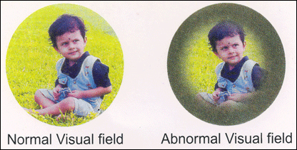
Automated Perimetry is an extremely sophisticated computerized procedure meant to test your field of vision. Since it is completely automated & computerized, the results are precise and accurate.
Why do we need to perform this test?
It is performed to test the field of vision in the following cases :
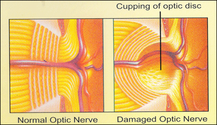
Glaucoma suspect/ Ocular hypertension to rule out presence of field defects. Glaucoma is an eye disorder damaging the optic nerve due to increase in the intraocular pressure. It is characterized by irreversible, progressive field loss.
Established glaucoma to look for progression of field defects.
Many neurological (brain) disorders.
Drug-induced / traumatic optic neuropathies & / or maculopathies.
This test can have an impact on further treatment of the above disorders. Hence, it is an extremely important diagnostic tool.
How do we perform test?
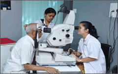
Automated Perimetry is an outpatient procedure taking 30-45 minutes. You will be made to sit in front of a computerized machine in a dimly lit room. Each eye will be tested separately. Glasses of your power will be inserted in the machine if required to give your clear vision. You will be asked to focus on a central light. A switch will be placed in your hand. After the initial settings, when the test begins, lights of varying intensity will be shone all around the machine in a random fashion. You will be asked to press the switch once immediately upon seeing the light. The switch should not be kept pressed for a long time.
Remember Please press the switch only if you see the light from the sides, but do not turn your eyes to look at them.
Some of the lights shown might be too dim for you to appreciate. But you need not worry about them. Visual field defects in glaucoma.
A technician will be guiding you through the procedure. In case of any watering of eyes or any other problem during the procedure you may either pause the test by keeping the switch pressed or tell the technician about it.
Throughout the procedure, you will be required to remain alert and attentive to the light stimuli. Excessive movement of the eyes and the head might cause significant errors in the report.
You need not come fasting for the test.
How often do we need to do the test?
Depending upon the patient’s eye condition, we may need to repeat the test every 6 months to 1 yr. This is to detect the progression of field defects and alter the treatment accordingly.
What is FFA?
Fluorescein Angiography is a dynamic study of the retino-choroidal status following administration of the eye, sodium fluorescein. This investigation forms an important armamentarium in the diagnosis & management of innumerable retinal disorders.
Frequently Asked Questions
Sequential fluorescein angiography has contributed greatly in understanding the natural course of numerous retinal disorders. Fluorescein Angiography is not only a diagnostic aid but also determines important decisions as in diabetic maculopathy. Indeed, in some disorders, a proper record & maintenance of sequential angiograms may be important as the maintenance of the ECG in a patient with schematic heart disease. It is also a useful aid to identify those patients at risk for developing further complications like neovascularization in whom early laser photocoagulation may hence be considered. Comparison with previous fluorescein angiograms is also often the basis on which laser augmentation is undertaken.
Some common indications of FFA are :
Diabetic Retinopathy
Hypertensive Retinopathy
Retinal Venous occlusions
Age related macular degeneration
Cystoids macular edema
Macular holes / macular dystrophies
This investigative procedure comprises of injecting a dye Fluorescein into one of your veins in the arm and taking rapid serial photographs of its passage within the inner structure of the eye the retina and choroids using a Fundus camera with appropriate filters.
The information obtained from a study of this procedure aids your doctor in making a diagnosis, planning your treatment or assessing the results of treatment particularly photo-coagulation.
There is no discomfort from this test apart from the needle prick and the flash of the camera which is harmless. You may have Nausea (sensation of vomiting) a minute or so after the injection. This usually passes off in about 30 seconds. Remaining calm and breathing deeply helps in overcoming this difficulty. You are advised to be an empty stomach 3 hours prior to this test. Your usual diet can be taken after the procedure Fluorescein is a highly non toxic drug. Rarely, it produces an allergic reaction which responds rapidly to appropriate medication. Serious life threatening reactions are exceptionally rare but can however occur. The skin and urine stain yellow for about 36 hours and is of no consequence.
Kindly read this and ask your doubts before fixing the test.
Patient is requested not to take anything by mouth for at least 3 hours prior to the test. This is to avoid vomiting. He /She are requested to take a light breakfast at 8.00 a.m. if asked to report at 10.00 a.m. for this test to prevent hypoglycemia.
He / She should take the usual medication for diabetes, hypertension or for any other systemic problems.
He / She should take the usual medication for diabetes, hypertension or for any other systemic problems.
The test will be performed � only if the B.P. is within normal limits. If it high (Diastolic more than 100 mm hg.) the test will be done after B.P. is normal, hence please check with your physician if your B.P. is high.
If patient is allergic to any drug, he should inform the doctor before the procedure.
Patients may have to wait for 1-2 hours because photography requires excellent dilation.
Diabetic patients are required to bring biscuits or glucose to take immediately after completion of the test to avoid hypoglycemia.
Kindly inform the doctor if the patient is pregnant, as it is preferable to avoid this test during first three months of pregnancy.
Patient has to be accompanied by an adult attendant.
Your FFA test has been scheduled on at
Please report at
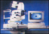
Optical Coherence Tomography(OCT)
Quick, safe and informative this new retina scanner is a glimpse into the future patient care.


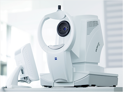
zies cirrus 5000 OCT machine IOL Master 700
Mrs. Bapat, your retina biopsy is complete. After examining a slice of your inner eye under a high-powered microscope, we are able to determine your vision problem and we never even touched your eye! Sounds space-aged? Not any more! Breakthroughs in ophthalmic imaging are making the retina’s inner workings transparent to a degree unimaginable even a few years ago. It’s called Optical Coherence Tomography, OCT for short. OCT is a non-invasive technology used for imaging the retina, the multi-layered sensory tissue lining the back of the eye. OCT, the first instrument used to see cross-sectional images of retina, is revolutionizing the early detection and treatment of eye conditions such as macular abnormalities & optic nerve damage.
How many ophthalmic imaging tests can claim the following?

Non-invasive
Non-contact
No radiation
Painless
Fast, it can provide quick, reproducible, high definition images.
Reliable and sensitive up to 5 microns (1 micron is a thousandth of a mm)
Virtually no set-up time, such as obtaining informed consent, installing film (old days), pre-treatment with antibiotics, etc.
Cirrus High Definition OCT
Cirrus High definition OCT is more advanced than its older model the stratus OCT in the following ways:
Faster-100 times more Ascans per second
More detailed-65 times higher resolution
More accurate-because of reduced artefacts
3-D imaging is possible
How does OCT work?
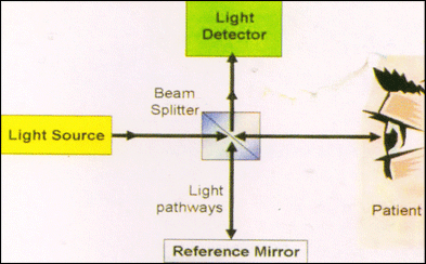
Rays of light provide 2 and 3-dimensional imaging of tissues at histological level.
The OCT machine has 3 modules:
OCT macula:
OCT macular scan report literally is like an Optical Biopsy of the 10 Retinal Layersng
OCT Macula :
Examines the retina and its sub-layers for structural integrity
Macular Atrophy
Macular edema
Macular traction
Macular holes
Sub retinal fluid
RPE irregularity
Clinical indications for macular/retinal OCT are :
Diagnosis of many retinal conditions like: Diabetic retinopathy esp maculopathy when changes are not yet evident on fundoscopy or on fluoroscein angiography.
Age related macular degeneration (ARMD)
Post cataract surgery Cystoid macular edema (CME)
Central serous retinopathy (CSR)
Macular holes
Epiretinal membranes
Pigment epithelial on neurosensory detachments at the macula
To monitor progression
To aid in treatment planning
To monitor response to therapy
OCT Optic disc for early diagnosis of glaucoma
Helps to measure the actual peripapillary retinal nerve fibre layer (RNFL) thickness. Studies have shown that the earliest signs of glaucoma are the thinning of the RNFL layer even before optic disc cupping is seen.
Hence, Extremely useful to detect glaucoma in its earlier stages even before Perimetry can detect field defects.
Useful in moniting glaucoma suspects or high risk patients for earliest signs of glaucoma progression.
OCT for anterior segment for examining cornea & anterior chamber angle structures