Keratoconus: Symptoms, Diagnosis And Treatments
Understanding Keratoconus symptoms causes and diagnosis in detail
- Keratoconus Symptoms Causes Diagnosis
- Keratoconus Prevention
- Keratoconus Early Diagnosis
Keratoconus is a progressive eye condition where the cornea thins and gradually bulges outward into a cone shape, leading to distorted vision. Let us understand keratoconus symptoms, causes diagnosis in detail:

What Are The Early Signs/Symptoms Of Keratoconus?
Keratoconus symptoms /early signs may include:
- Blurred or Distorted Vision: This is one of the most common early symptoms. Patients may notice that their vision becomes blurry or distorted, particularly when looking at objects from a distance.
- Increased Sensitivity to Light: Some individuals may experience heightened sensitivity to light (photophobia), causing discomfort or glare in bright light conditions.
- Frequent Changes in Eyeglass Prescription: Because keratoconus causes changes in the shape of the cornea, individuals may find that their eyeglass prescription changes frequently, often needing stronger or different prescriptions.
- Astigmatism: Astigmatism is a common symptom of keratoconus, where the cornea becomes irregularly shaped, leading to blurred or distorted vision at all distances.
- Difficulty Seeing at Night: Patients with keratoconus may experience difficulties seeing clearly at night, often due to increased glare or halos around lights.
- Eye Irritation or Discomfort: Some individuals may experience eye irritation, itchiness, or a sensation of foreign objects in the eye.
- Frequent Rubbing of the Eyes: Continuous rubbing of the eyes can exacerbate keratoconus symptoms. It may also be a sign that the eyes are experiencing discomfort or visual disturbances.

What Are The Causes Of Keratoconus?
The exact keratoconus cause is not fully understood, but it is believed to involve a combination of genetic, environmental, and biochemical factors. Some of the main factors thought to contribute to the development of keratoconus include:
- Genetics: There is evidence to suggest that genetics play a significant role in the development of keratoconus. The condition often runs in families, and individuals with a family history of keratoconus are at a higher risk of developing it themselves.
- Corneal Structure: Abnormalities in the structure and composition of the cornea may contribute to the development of keratoconus. This includes thinning of the cornea, which weakens its structural integrity and allows it to bulge outward into a cone shape.
- Eye Rubbing: Continuous rubbing of the eyes is believed to be a risk factor for keratoconus, as it can exacerbate corneal thinning and deformation. Rubbing the eyes may be more common in individuals with allergies or other eye conditions that cause itching or irritation.
- Eye Trauma: Trauma to the eye, such as excessive eye rubbing, eye injuries, or wearing poorly fitted contact lenses, may increase the risk of developing keratoconus.
- Biochemical Imbalances: Some researchers believe that biochemical imbalances within the cornea, such as increased levels of enzymes called proteolytic enzymes, may contribute to the weakening and thinning of the cornea in keratoconus.

How Do You Diagnose Early Keratoconus?
Diagnosing early keratoconus typically involves a comprehensive eye examination by an eye care professional. Some of the key diagnostic tests and procedures used to detect early keratoconus include:
- Corneal Topography: Corneal topography is a non-invasive imaging technique that maps the curvature and shape of the cornea’s surface. It can detect subtle irregularities in the cornea’s shape characteristic of keratoconus, even before symptoms become apparent.
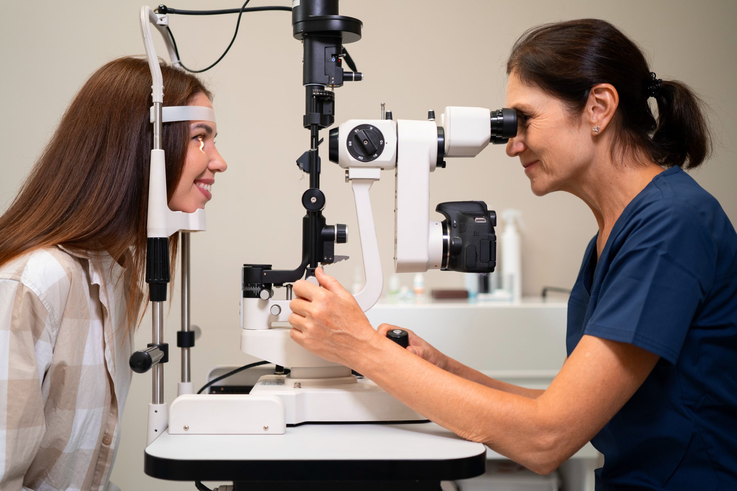
- Slit-Lamp Examination: During a slit-lamp examination, the eye care professional examines the cornea using a special microscope called a slit lamp. This allows them to assess the cornea’s clarity, thickness, and any structural changes indicative of keratoconus.
- Refraction Test: A refraction test measures the eye’s ability to focus light and determines the need for corrective lenses. Individuals with keratoconus often have irregular astigmatism, which can be detected during this test.
- Visual Acuity Testing: Visual acuity testing assesses the clarity of vision at various distances using an eye chart. Early keratoconus may cause blurry or distorted vision, which can be detected through changes in visual acuity.
- Pachymetry: Pachymetry measures the thickness of the cornea. Individuals with keratoconus often have thinner corneas compared to those without the condition.
- Ocular Surface Examination: An examination of the ocular surface may reveal signs of eye rubbing, allergies, or other conditions that can contribute to keratoconus or exacerbate its symptoms.
- Family History and Symptoms: In addition to objective tests, the eye care professional will inquire about any family history of keratoconus and ask about symptoms such as blurry vision, frequent changes in prescription, or sensitivity to light.

Conclusion:
Early diagnosis of keratoconus is crucial for initiating appropriate management and treatment to help slow its progression and preserve vision. If keratoconus is suspected, further evaluation by a cornea specialist may be recommended for confirmation and to discuss treatment options.
Recent Post
Best Lasik Eye Surgery: It’s Procedure and Benefits
Understanding Best Lasik Eye Surgery What is LASIK? How Does LASIK Working? Benefits of LASIK Eye Surgery Procedure of Best Lasik…
Eye Care Routine for Maintaining Good Eye Health During Monsoon
Maintaining Good Eye Heath During Monsoon Common Eye Allergies Eye Care Tips To Prevent Your Eyes From Damage Maintaining good eye…
Dry Eye: The importance of tears in maintaining healthy eyes
Dry Eye: The importance of tears in maintaining healthy eyes Understanding Dry Eye Managing Dry Eye Syndrome Dry Eye: The importance…


